Cart (0 Items)
Your cart is currently empty.
View ProductsIt looks like you are visiting from outside the EU. Switch to the US version to see local pricing in USD and local shipping.
Switch to US ($)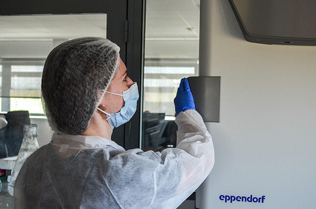
With 25+ years of experience and a track-record of more than 500 phage display projects, ProteoGenix can confidently guide you to your project’s success and offer the strongest guarantees of the market.
Discover why more than 9,000 researchers choose ProteoGenix.
GUARANTEED
If we do not provide, we screen 96 additional candidates for free
NO HIDDEN FEES
No IP contraints, no royalties
MAXIMUM DIVERSITY
Camelids (VHH), humans, dogs, rabbits and cats
PROPRIETARY TECHNOLOGY
Including world’s first human cancer & autoimmune libraries
PHD GUIDANCE
Your dedicate PhD Expert will guide you and provide advice throughout your entire project
FULLY CUSTOM
To ensure success, the experimental strategy is adapted to your project and antigen
Whatever your research focus, we match the phage display service to your application. You can choose from highly targeted libraries, including our Human Cancer Library covering 12 cancer types and our Autoimmune Library spanning 12 diseases such as Sjögren’s syndrome and Crohn’s disease. For animal health projects, we provide species-specific libraries for cat, dog, pig, camelid, mouse, and rabbit, so you get binders that fit your model from the start.

If your project requires human antibodies, our phage display service is the most direct route. Thanks to our broad portfolio of human libraries, we generate fully human binders upfront, with no humanization step, so you can move faster into development with sequences already aligned to clinical needs.

Working on a complex target such as GPCRs, virus-like particles, or MHC–peptide complexes? We can support you with confidence. With more than 20 years of phage display expertise, ProteoGenix has a proven track record on challenging antibody projects, delivering high-quality binders even when targets are difficult to express, unstable, or highly conformational.

Thanks to our expertise and broad technology panel, we also provide phage display for special formats such as VHH. Using our dedicated VHH library, we generate stable single-domain antibodies that access hard-to-reach epitopes and support next-generation therapeutic designs.

Get antibodies in as little as 4 weeks with a workflow built around your timeline. Our pre-built libraries accelerate selection without compromising quality. We adapt our phage display services to your deadlines so you receive validated binders when you need them.
Human – Donors who recovered from COVID-19.
Application: Ideal for COVID-19 antibody generation
Format: ScFv
Size: 1.19 x 10¹⁰ clones
Human – PBMCs & BMMCs from Human Donors. 18 different cancers.
Application: Ideal for custom cancer immunotherapy development
Format: ScFv & Fab
Size: 6.81×10¹⁰ ScFv & 6.72×10¹⁰ Fab
Human – PBMCs & BMMCs from Human Donors. 12 different autoimmune diseases.
Application: Ideal for custom autoimmune disease therapy development
Format: ScFv & Fab
Size: 1.08×10¹¹ ScFv & 1.06×10¹¹ Fab
We create your custom library with an immunization protocol adapted to your project.
Application: Research, Diagnostic, Therapeutic, Animal Health
Format: Your choice


Tyr Pharma, USA
Senior scientist at aTyr Pharma
“All three of the antibodies you delivered were reformatted into IgGs and worked well. Interestingly two were d1 binders, and one was a d1/d2 binder when we domain mapped them here. They were good “protein X” blocking antibodies. But none were “protein Y” blockers. They express reasonably well. Overall we are quite happy with them.”
Nanyang Technological University, Singapore
Alvin Chew, Research Fellow
“ProteoGenix has been generous and sincere in supporting an academic laboratory like ours. They were able to identify several antibody sequences via phage display against a protein antigen and they screened twice more phages than initially planned to help us succeed in our project. ProteoGenix team was reactive and diligent in their replies and services.”

Brown University, USA
Prof Jonathan Kurtis, MD, PhD, Chair, Department of Pathology and Laboratory Medicine
“I requested ProteoGenix’s services for the generation of antibodies against a malarial protein by phage display. Their naive human library, which is comprised of 368 donors, is a unique resource which was one of the criteria I used to select their company. They optimized their standard screening strategy to be able to identify two specific clones, that they then expressed with very high yields in their proprietary XtenCHOTM cell line. I highly appreciated their level of expertise and their commitment to go beyond expectations, and I would recommend them for their reactivity and the high quality of their services.”
UiT the Arctic University of Norway, Tromsø, Norway
Dr Lorena Arranz, Group Leader – NCMM Associate Investigator – SFF Center Director
“We requested ProteoGenix’s services for the development of a monoclonal antibody for flow cytometry. They were able to identify three candidates that all bound well and specifically to our antigen. The best one was validated in our final application and the results were published in an international journal. We were overall satisfied with the quality of the final product and its results.”
National Cancer Centre, Singapore
Prof Johnny Ong Chin-Ann, MD, PhD, Group leader
“We have been working together with ProteoGenix since 2020. Using phage display, they helped our team to develop several antibodies with effective blocking effects. We are pleased with their services and look forward to continue working together with them as we advance our research and bring the biologics to clinical stage.”
University College London, UK
Markella Ponticos – Professor
“Our collaboration with ProteoGenix focused on the generation of specific monoclonal antibodies directed towards a mammalian cell surface protein. The antibodies developed exhibited very high affinity and performed in cell-based functional assays. This data propelled us towards advancing into pre-clinical in vivo and human work for the refinement and development of a potent biologic reagent. The invaluable technical and scientific guidance along with the expertise and support provided by our liaison at ProteoGenix played a pivotal role in the collaboration and advancing our translational research.”