

Cart (0 Items)
Your cart is currently empty.
View ProductsIt looks like you are visiting from outside the EU. Switch to the US version to see local pricing in USD and local shipping.
Switch to US ($) Antibody-drug conjugates
 Thomas Meyer
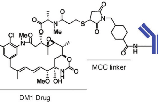
Antibody-drug conjugates (ADCs) are highly efficient targeted delivery systems for small cytotoxic agents. Covalently bound to the antibody via cleavable or non-cleavable linkers, the payloads of ADCs are the sole bioactive components of these biotherapeutics. However, the complexity of these systems often leads to substantial heterogeneity, warranting an extensive characterization of each of the individual components and the conjugated molecule as a whole before they can be considered suitable for clinical applications.
The drug-to-antibody ratio (DAR) and the drug load or DAR distribution are two of the major critical properties of ADCs. The DAR value is defined as the average number of drug molecules conjugated to an antibody. In contrast, drug load or DAR distribution is defined as the stoichiometric distribution of the number of conjugated drugs present on the antibody carrier. DAR values dictate the efficacy and safety of ADCs. High DAR may hinder the pharmacokinetic properties of ADCs, while low DAR significantly limits the potency of these therapeutics. Ideally, DAR distribution values should be between 2-4.
Several methods have been developed to measure these properties with increasing accuracy. However, the inherent heterogeneity of these systems and the presence of complex post-translational modifications in the antibody carrier make this characterization analytically challenging.
Currently, these properties are measured using the following methods:
UV/Vis spectroscopy is the simplest methodology used for ADC characterization. Traditionally used to determine protein concentration, this method relies on measuring the absorbance at different wavelengths (λ). The only prerequisites for this method are: (i) the payload must have a UV/Vis chromophore; (ii) the antibody and payload should have distinctive wavelengths of maximum absorbance (λmax values, the former usually at 280 nm); (iii) the drug must not interfere with the absorption spectra of the antibody and vice-versa.
If the ADC complies with all prerequisites, the concentration of both components can be calculated based on absorbance values and extinction coefficients. Subsequently, the average DAR can be estimated simply by dividing the average drug concentration by the average antibody concentration.
Although this methodology is routinely used to estimate the DAR of ADCs, it presents important limitations. For instance, the presence of free drug in an ADC sample can lead to the overestimation of DAR values. Moreover, UV/Vis spectroscopy does not provide any information regarding the drug load distribution, an equally important property. For this reason, this method is reserved for routine quality control operations and is rarely used during the early development of new ADCs.
Cathepsin B is a lysosomal enzyme responsible for cleaving ADC constructs with peptide linkers. The development of an enzymatic method based on cathepsin B for DAR determination is recent. The methodology involves a series of treatments with several enzymes and a reductant to allow the complete release of conjugated drugs. The quantification of the free drug after treatment is subsequently carried out by chromatographic separation.
The ADC is initially pretreated with IdeS protease to yield F(ab′)2 and Fc fragments followed by reduction with 2-mercaptoethylamine (2-MEA) to allow further fragmentation into Fdʹ, Fc, and Lc. This complex pretreatment ensures better access to conjugation sites and more efficient cleavage of the payload. Following these steps, digestion with cathepsin B is carried out and subsequent separation of protein component is performed by RP-HPLC-UV.
Because both components are separated before drug quantification, this method overcomes the most common limitation imposed by UV/Vis methods – the need for distinct λmax values for the drug and antibody component. Additionally, this method is also considered more precise than mass spectrometry where the mass signal can often be affected by hydrophobicity and net protein charge, and hydrophobic interaction chromatography (HIC) that may suffer from low-resolution efficiency when analyzing randomly conjugated ADCs.
The most important limitation of cathepsin B-based methods is the inability to discern drug load distribution. Moreover, due to the complexity of the method, its implementation for quality control purposes remains challenging. For this reason, the method remains more useful in early development or antibody research contexts.
Capillary electrophoresis (CE) is characterized by simple instrumentation, miniaturized format, efficient and rapid separation, low sample input, and high resolution. Different CE methods have been tested and implemented for the characterization of ADCs including capillary gel electrophoresis (CE-SDS), capillary isoelectric focusing (cIEF), imaged cIEF (iCIEF), capillary zone electrophoresis (CZE), and capillary electrophoresis–mass spectrometry (CE–MS). All these methodologies offer different levels of characterization for ADCs such as isoform, glycol-profiling, and post-translational modifications, among others.
Like cathepsin B-based methods, CE methodologies have only been recently developed. For this reason, little literature is available to support them. However, among these methodologies, iCIEF has consistently led to promising results. The technique allows the separation of ADC charge variants based on their isoelectric point (pI). In this way, unconjugated antibodies and different ADC species with different drug loads can be separated efficiently, helping to monitor batch-to-batch consistency, drug load distribution, and unconjugated antibodies. Its sole drawback is that the technique has only been tested on lysine-based conjugated ADCs. For this reason, it is still unclear if it can be applied to other conjugation chemistries.
Because detailed DAR analysis depends heavily on conjugation chemistry and molecular format, it is often best interpreted within broader antibody engineering services and ADC optimization workflows.
Hydrophobic interaction chromatography (HIC) works by running an inverse salt gradient on a hydrophobic stationary phase. The stationary phase retains and separates sample components based on their hydrophobicity, and because the method uses mild conditions, the native conformation of complex protein species is preserved during the analysis. For this reason, HIC methodologies are useful to measure average DAR and determine drug load distribution in ADCs.
The use of mild conditions has made this technique extremely convenient when analyzing non-covalent protein conjugates such as cysteine-linked ADCs. These conjugates are often dissociated during harsher chromatographic methods such as RPLC (reverse-phase liquid chromatography), making HIC the technique of reference for the detailed DAR analysis of these biopharmaceuticals.
HIC techniques are considerably less efficient at analyzing lysine-based and site-specific conjugated ADCs. Moreover, the ability of the technique to separate unconjugated molecules remains limited as these species are only partially resolved. However, due to the recent development of new columns and separation protocols, HIC continues to be the gold standard for the drug load distribution analysis of cysteine-linked ADCs.
Reversed-phase high-performance liquid chromatography (RP-HPLC) is widely used to characterize pharmaceutical proteins. The advantage of RP-HPLC over HIC-based characterization is the compatibility of this method with mass spectrometry due to the volatility of the mobile phase. In contrast, HIC uses a salt gradient incompatible with MS studies. RP-HPLC or simply RPLC (reverse-phase liquid chromatography), has a successful track record as the method of choice to analyze average DAR, drug load distribution, unconjugated antibodies, and free drug-linker species.
For cysteine-linked ADCs, RPLC is the most suitable alternative to HIC for detailed DAR analysis. However, one of the major drawbacks of this method derives from the harsh analytical conditions known to disrupt the native conformation of cysteine-linked ADCs, often leading to the dissociation of light and heavy chains. To overcome this limitation, ADCs are frequently pre-treated with reductants prior to analysis. In this case, the light and heavy chains are more easily separated and individually assessed. Alternatively, ADCs can also be pre-treated with IdeS and chemically reduced to generate Fc, LC, and Fd fragments.
Finding suitable chromatographic conditions remains challenging independently of the protocol used for ADC analysis in RPLC. And for lysine-linked ADCs, the resolution provided by RPLC separation is often insufficient to discriminate between intact DAR species with different drug loads. For this reason, this methodology is effective for average DAR determination but challenging when analyzing drug load distribution in lysine-linked ADCs. In contrast, site-specific conjugated ADCs are easily analyzed both by HPLC or HIC-based methodologies.
In general, independently of the conjugation chemistry (cysteine, lysine, or site-specific), RPLC serves as a robust method for quality control of average DAR and is extremely useful to detect the presence of impurities. Moreover, RPLC methods can be used to estimate ADC stability and quantify free drug molecules, making them useful for the extensive early characterization of novel ADCs and optimization of their production process.
DAR analysis is most powerful when it is integrated into a broader ADC development workflow. ProteoGenix offers antibody-drug conjugate development services with a full analytical suite covering DAR, DAR distribution, drug location, % free drug, and standard antibody QC measurements, combined with 20+ years of expertise and 5 ADCs in clinical trials. For conjugation itself, ProteoGenix reports >90% DAR homogeneity at the specified ratio, which helps reduce ADC toxicity and improve consistency. This makes our platform particularly relevant for teams working on DAR control, DAR distribution optimization, and analytical characterization during early development and preclinical preparation.
Mass spectrometry (MS) analysis depends on the ionization efficiency of the analyte and comprises several powerful and high-resolution techniques. Currently available MS spectrometers for the analysis of large biomolecules use electrospray ionization (ESI) coupled with time-of-flight (TOF) or Orbitrap, which help different ADC species with high resolution in reducing or non-reducing conditions.
Due to the sensitivity of these methods, it is often advisable to pre-treat the ADCs with PNGase F to eliminate the heterogeneity caused by N-glycans, frequently responsible for masking or interfering with MS analysis. Popular alternatives include the reduction of ADCs into heavy and light chains, enzymatic fragmentation of the antibody carrier, or a combination of the two. Additionally, for lysine-linked ADCs which usually have a particularly low ionization efficiency, researchers have successfully removed the hydrophobic portion of the payload to successfully increase detection sensitivity.
Pre-treatments typically allow a more detailed structural analysis of ADCs including the detection of low abundance ADC species with high accuracy and sensitivity.
All the different methodologies covered in this article should be seen as complementary. In fact, the comparison of results obtained using different methodologies contributes to a more accurate fingerprint of the ADC molecule in regards to its average DAR, drug load distribution, unconjugated antibody content, and free payload-linker content. Moreover, the performance and accuracy of most of these techniques is heavily dependent on conjugation chemistry. For this reason, several methods should be used during early development to ensure a proper and extensive characterization of these biopharmaceuticals.
Although MS-based methodologies continue to be the best in terms of resolution, their implementation for routine quality control is not often feasible. For this reason, alternative methods such as HIC or CE should often be preferred to expedite ADC production and quality control.
Join our email list to receive exclusive content featuring the most interesting industry and research news, biologics development tips pieced together by experts, res, company news, and exclusive limited-offers. Join a community of 80,000 subscribers and save up to 30% on your first order.